FIGURE 5.
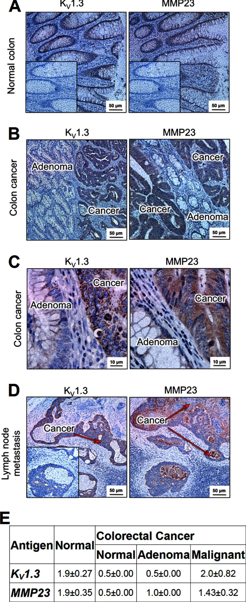
Expression of KV1.3 and MMP23 in human colon. A, immunohistochemistry of normal human colon (at 20×) showing KV1.3 (left) and MMP23 (right) in epithelial cells. Inset, the same isotype control was used for both KV1.3 and MMP23; polyclonal rabbit IgG was used in place of the primary rabbit anti-KV1.3 or rabbit anti-MMP23 antibodies. B, KV1.3 (left, 20×) and MMP23 (right, 20×) expression in colorectal cancer. Arrows indicate adenoma (yellow) and KV1.3- and MMP23-expressing malignant colorectal epithelium (red). C, higher magnification images of colorectal cancer showing KV1.3 (left, 100×) and MMP23 (right, 100×). Note intracellular staining of both proteins. D, KV1.3 (left, 20×) and MMP23 (right, 20×) in metastatic colorectal epithelium in lymph nodes. The red arrows highlight the KV1.3- and MMP23-expressing metastatic colorectal epithelium. The surrounding lymph node is not stained. E, scoring of staining intensity on a scale of 1–3 of KV1.3 and MMP23 in epithelium from normal colon and from three regions of colonic tissue from patients with colorectal cancer: normal-looking crypts, adenoma, and malignant epithelium.

