FIGURE 1.
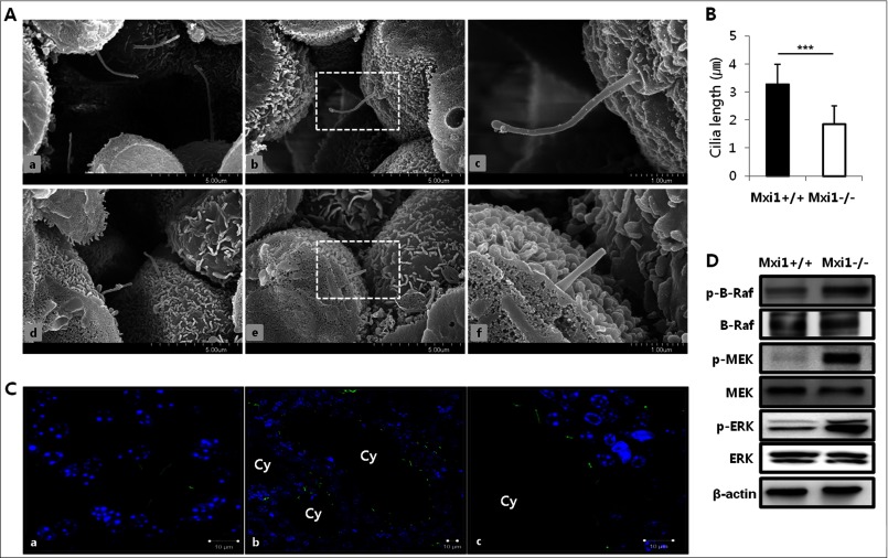
Identification of primary cilium in kidney of Mxi1-deficient mice. A, representative photographs of scanning electron micrographs of kidneys from wild-type (a–c), aged 9 months, and Mxi1-deficient kidney (d–f) as age-matched controls. B, comparison of the length of cilium in Mxi1-deficient kidney with wild-type kidney. The length of cilium was quantified by measuring from ciliary tip to basal body (basal body is bottom and protruding region of cilia) using the scale bar indicated in the figure. The graph shows mean ± S.D. (error bars) of three independent experiments. The one-tailed p value is <0.0001, considered extremely significant (***). C, representative photographs of anti-acetylated α-tubulin-stained Mxi1-deficient kidney (b and c) and wild-type kidney (a) (anti-acetylated α-tubulin staining (green), DAPI staining (blue), Cy, cyst. Original magnification, ×3000. Scale bars, 10 μm. D, Western blot of proteins related to B-Raf/MEK/ERK pathway in Mxi1-deficient kidney and wild-type. β-Actin was used as a loading control.

