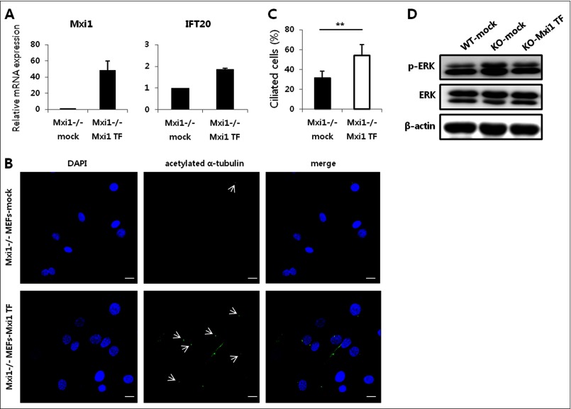
FIGURE 4.
Transient overexpression of Mxi1 has an effect on ciliary phenotype of Mxi1−/− MEFs. A, verification of Mxi1 and Ift20 mRNA expression in Mxi1−/− and Mxi1 transiently overexpressed Mxi1−/− MEFs. The graphs show mean ± S.D. (error bars) in triplicate. B, representative photographs of anti-acetylated α-tubulin-stained Mxi1−/− and Mxi1 transiently overexpressed Mxi1−/− MEFs. White arrows indicates cilia stained with anti-acetylated α-tubulin. Original magnification, ×400. Scale bars, 20 μm. C, quantification of ciliated cells in Mxi1−/− and Mxi1 transiently overexpressed Mxi1−/− MEFs. To measure ciliated cells, the ratio of the number of cilia to the number of nuclei was calculated and multiplied by 100. The graph shows mean ± S.D. of three independent experiments. The one-tailed p value is 0.007, considered significant (**). D, Western blot of p-ERK and total ERK in Mxi1+/+, Mxi1−/−, and Mxi1 transiently overexpressed Mxi1−/− MEFs. β-Actin was used as a loading control.

