FIGURE 2.
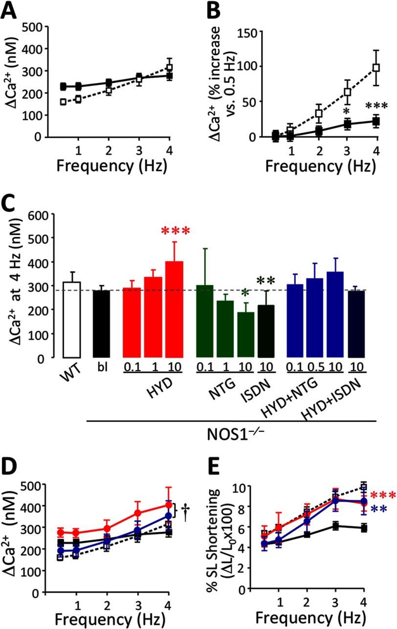
Intracellular Ca2+ transient (Δ[Ca2+]i) in NOS1−/− cardiomyocytes. A, Δ[Ca2+]i (nm) in NOS1−/− (■; n = 40–50 cells) compared with WT (□; n = 32–40 cells). B, Δ[Ca2+]i in NOS1−/− (■) and WT (□) CMs, expressed as % increase versus 0.5 Hz. C, Δ[Ca2+]i at 4 Hz in WT control or NOS1−/− CMs in the absence (bl) or presence of HYD (0.1, 1 or 10 μm), NTG (0.1, 1 and 10 μm), ISDN (10 μm), and a combination (HYD + NTG; 0.1, 0.5, and 10 μm each; and HYD + ISDN; 10 μm each). D, ΔCa2+ frequency response in WT CMs (□) under baseline conditions or NOS1−/− baseline (■) or treated with 10 μm HYD (red circle) or 10 μm HYD + NTG (blue circle). E, FFR in CMs under the same conditions as mentioned above. The combination induces a significant increase in FFR but does not change ΔCa2+ significantly compared with NOS1−/− CMs. However, the contractile reserve is restored. *, p < 0.05; **, p < 0.01; and ***, p < 0.001 versus NOS1−/− control; †, p < 0.05 versus HYD alone; two-way ANOVA.

