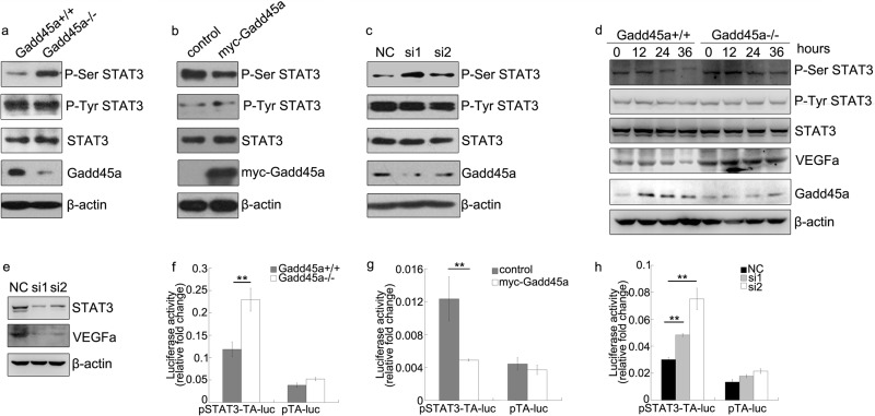
FIGURE 6.
Gadd45a defect leads to up-regulated P-Ser STAT3 and transcriptional activity. a, Gadd45a+/+ and Gadd45a−/− MEFs were harvested and prepared for Western blot analysis with P-Ser STAT3, P-Tyr STAT3, and Gadd45a antibodies. b and c, HeLa cells were transiently transfected with myc-Gadd45a vector or siRNAs for Gadd45a. The variation of the phosphorylation of STAT3 and total STAT3 was analyzed by immunoblots 36 h after transfection. Myc-Gadd45a was detected with myc antibody. NC was a negative control of siRNA. d, Gadd45a+/+ and Gadd45a−/− MEFs were collected for an immunoblotting assay after serum starvation for different time points, which are marked at the top of the panels. β-actin was used as the loading control. e, VEGFa expression was further evaluated after STAT3 knocked down in HeLa cells. f, for the luciferase assay, pSTAT3-TA-luc and internal control plasmid pRL-SV40 were transiently cotransfected into Gadd45a+/+ and Gadd45a−/− MEFs. The luciferase activity was normalized against the internal control. The data were presented as mean ± S.E. n = 3. g and h, the STAT3 reporter luciferase assay was performed after pSTAT3-TA-luc and pRL-SV40 were cotransfected with myc-Gadd45a vector or siRNAs for Gadd45a into HeLa cells. The relative luciferase activity was presented as mean ± S.E. n = 3. **, p < 0.01).

