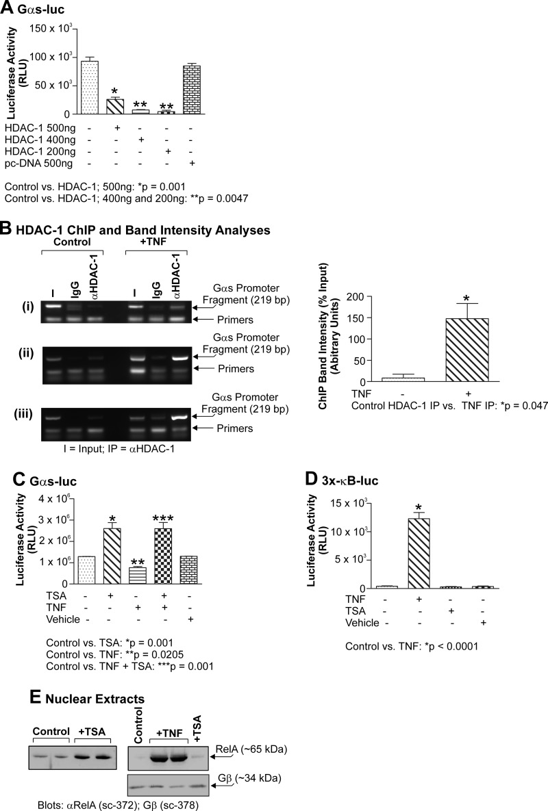
FIGURE 7.
Gαs-luc activity is repressed by HDAC-1 but induced by TSA. Primary myometrial cultures were transfected with 200 ng of Gαs-luc and 200, 400, or 500 ng of HDAC-1. Cells were harvested after 24 h, and luciferase activity was quantitated. HDAC-1 strongly repressed Gαs expression at all doses and maximally repressed Gαs expression at 200 ng (A; p = 0.0047). Primary cultures of myometrial cells were stimulated with TNF (10 ng/ml) for 24 h and subjected to the ChIP assay with anti-HDAC-1 antiserum. Data were compared using a paired, two-tailed t test, and results are expressed as the mean ± S.E. (error bars); p < 0.05 was considered statistically significant. TNF was seen to induce an increase in HDAC-1 occupancy of the endogenous Gαs promoter (B, panels i–iii; p = 0.047). Primary myometrial cultures were transfected with 200 ng of Gαs-luc and then 24 h later stimulated with TNF (10 ng/ml) for 24 h. TSA (330 nm) was then added to the culture medium for 24 h. Cells were harvested, and luciferase activity was quantitated. Data were compared using one-way analysis of variance and analyzed further using Tukey's multiple comparison test, and results are expressed as the mean ± S.E. (error bars); p < 0.05 was considered statistically significant. TSA was seen to activate Gαs-luc activity and overcome TNF-induced Gαs-luc repression (C; p < 0.001). Primary myometrial cultures were transfected with 200 ng of Gαs-luc and then 24 h later stimulated with either TNF (10 ng/ml) for 24 h, TSA (330 nm) for 24 h, or TNF and then TSA. Only TNF activated the NF-κB-sensitive 3x-κB-luc reporter (D; p = 0.0001). Cultures of primary myometrial cells were stimulated with either TNF (10 ng/ml) for 24 h, TSA (330 nm) for 24 h, or TNF and then TSA. Nuclear extracts were subsequently prepared and probed for the expression of acetyl-RelA (Lys-310) and total RelA. No acetyl-RelA was detected in response to any stimulant (not shown). TSA, however, induced significant RelA nuclear localization, but this remained less than that seen with TNF (E, upper panels). Equal loading was confirmed by blotting for Gβ (E, lower panel). All experiments were performed three times, and results are expressed as the mean ± S.E. (error bars). RLU, relative light units. I, input; IP, immunoprecipitate.

