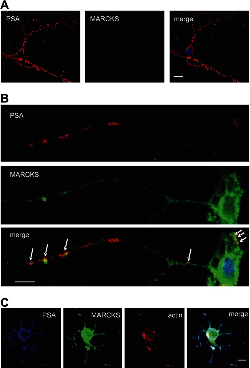
FIGURE 4.
Co-localization of MARCKS and PSA in cultured hippocampal neurons. A and B, cultured live hippocampal neurons (A) or fixed hippocampal neurons (B) were incubated with rabbit polyclonal MARCKS antibody and mouse monoclonal PSA antibody followed by incubation with corresponding fluorescent dye-labeled secondary antibodies. After fixation and permeabilization of cells, nuclei were stained with DAPI. Superimpositions of MARCKS and PSA staining show partial co-localization (seen in yellow) at the surface of cell bodies and processes. C, cultured live hippocampal neurons were incubated with mouse monoclonal PSA antibody followed by fixation and permeabilization of the cells and by incubation of the fixed cells with rabbit polyclonal MARCKS antibody, fluorescent dye-labeled secondary antibodies, and phalloidin. Superimpositions of MARCKS, PSA, and actin staining show partial co-localization (seen in white) at the surface of cell bodies and processes. The scale bar represents 5 μm.

