FIGURE 6.
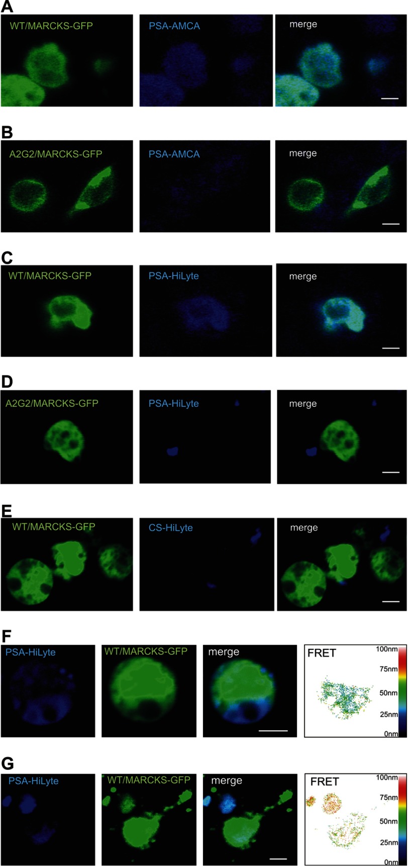
PSA and MARCKS interact at the cell membrane from opposite sides. CHO cells (A–F) or hippocampal neurons (G) were transfected with non-mutated MARCKS-GFP (A, C, and E–G) or with a non-myristoylatable MARCKS-GFP mutant (B and D). Colominic acid/PSA conjugated to the fluorescent dye aminomethylcoumarin (AMCA; A and B) or HiLyte Fluor 405 (C, D, F, and G) or chondroitin sulfate conjugated to HiLyte Fluor 405 (E) was applied to the transfected cells. A–G, images of representative cells from confocal microscopy and FRET analysis are shown. The scale bar represents 5 μm.

