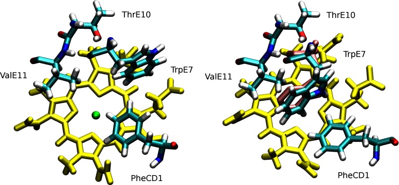
FIGURE 4.
Conformational switch of Trp-E7 from the outward conformation (left panel) to the inward conformation (right panel). Trp-E7, Phe-CD1, Thr-E10, and Val-E11 are depicted in cyan. In both panels, the E7 pathway is blocked by the bulky Trp side chain (see Fig. 3). Position of Trp-E7 in the crystal structure of the Trp-E7 mutant α subunit of recombinant human hemoglobin with wild-type β subunit was superimposed in violet (Protein Data Bank code 3NMM) (12).

