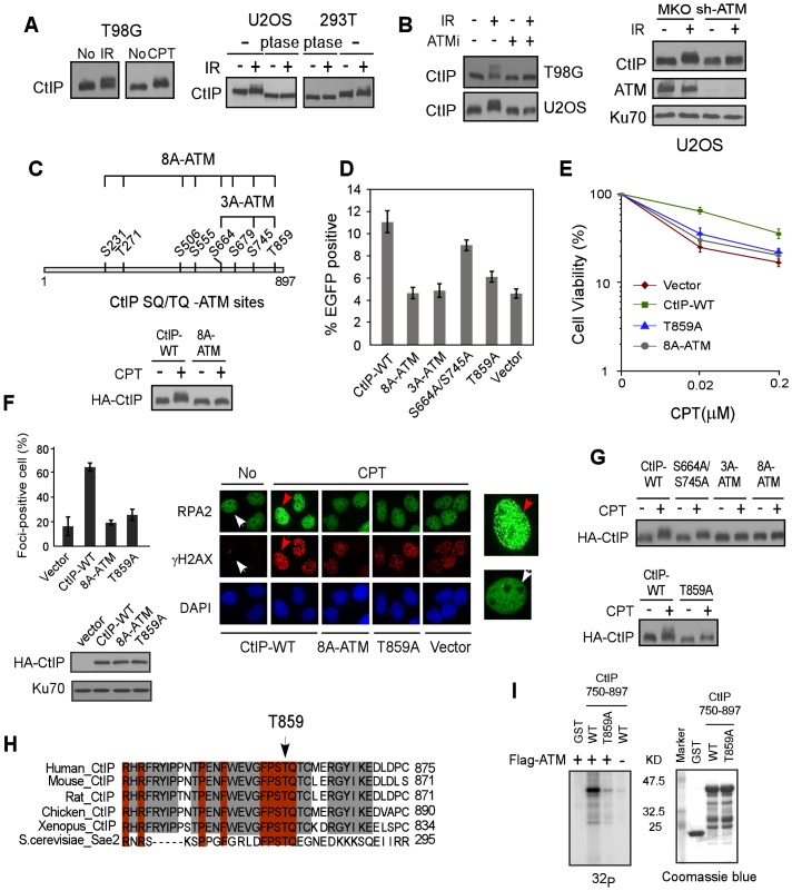
Figure 1. ATM is important for CtIP hyper-phosphorylation after DNA damage.
A. Indicated cell lines were treated with IR (10 Gy) or CPT (2 µM, 2 h) or No and lysed. Western blotting of CtIP was performed using cell lysates incubated with lambda phosphatase (ptase) or without ptase (-). B. U2OS and/or T98G cells were treated with ATM inhibitor (ATMi) KU-55933 (20 µM, 1 h) or infected with retroviruses encoding shRNAs for ATM or vector control MKO, treated with IR (10 Gy) and lysed 1 h later. Western blotting was performed with indicated antibodies. C. Schematic drawing of eight putative ATM consensus sites (SQ/TQ motifs) on CtIP. U2OS cells stably expressing HA-CtIP wild-type (WT) or HA-CtIP 8A-ATM mutant (all SQ/TQ sites mutated to AQ) with endogenous CtIP silenced by shRNAs and siRNA, were treated with CPT (2 µM, 2 h) followed by anti-HA Western blotting. D–F. EGFP-based HR assay, clonogenic survival and RPA foci formation assays were performed with U2OS cells stably expressing indicated CtIP variants, with endogenous CtIP silenced. Data shown represents the mean of three independent experiments; error bars, s.d. In panel F, indicated cells were treated with DMSO (No) or CPT (1 µM, 2 h) and fixed for immunostaining. The red and white arrows indicate representative RPA2 and γH2AX foci-positive and foci-negative cells, respectively, with RPA2-immunostained cells enlarged in right-side panels. The percentage of RPA2 foci-positive cells among γH2AX-positive cells for each sample is shown. G. Western blot of U2OS cells expressing indicated CtIP variants with endogenous CtIP silenced and treated with CPT (2 µM, 2 h). H. The C-terminus protein sequence of human CtIP was aligned with CtIP homologues from other indicated species, with newly identified conserved T859 site shown. I. ATM in vitro kinase assay was performed with purified GST-tagged CtIP fragments (residues 750–897), containing either WT or the T859A mutation. Coomassie Blue staining indicates protein loading.

