
Figure 6. Methylation protection of the vlsE G4-forming sequence.
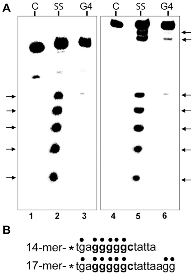
A) Autoradiogram of a 25% denaturing polyacrylamide gel of dimethylsulfate treated 14-mer and 17-mer oligonucleotides. 32P-labeled oligonucleotides were annealed in 200 mM KCl and treated with 0.5% dimethylsulfate. The methylated oligos were then subjected to electrophoresis in a 20% native polyacrylamide gel to separate the single stranded (SS) oligos from the G4-DNA. The free and the G4 bands were treated with 1 M piperidine to induce strand breaks at the methylated guanine residues and the cleavage products were resolved on a 25% denaturing polyacrylamide gel. C represents the control oligo which was not treated with DMS. The arrows correspond to the DMS-protected guanine residues. B) Sequence of the oligonucleotides used in this experiment. The dots show the position of G residues. The smallest cleavage fragment was run off the bottom of the gel.