
Figure 6. Δpbcax parasite ookinete conversion images.
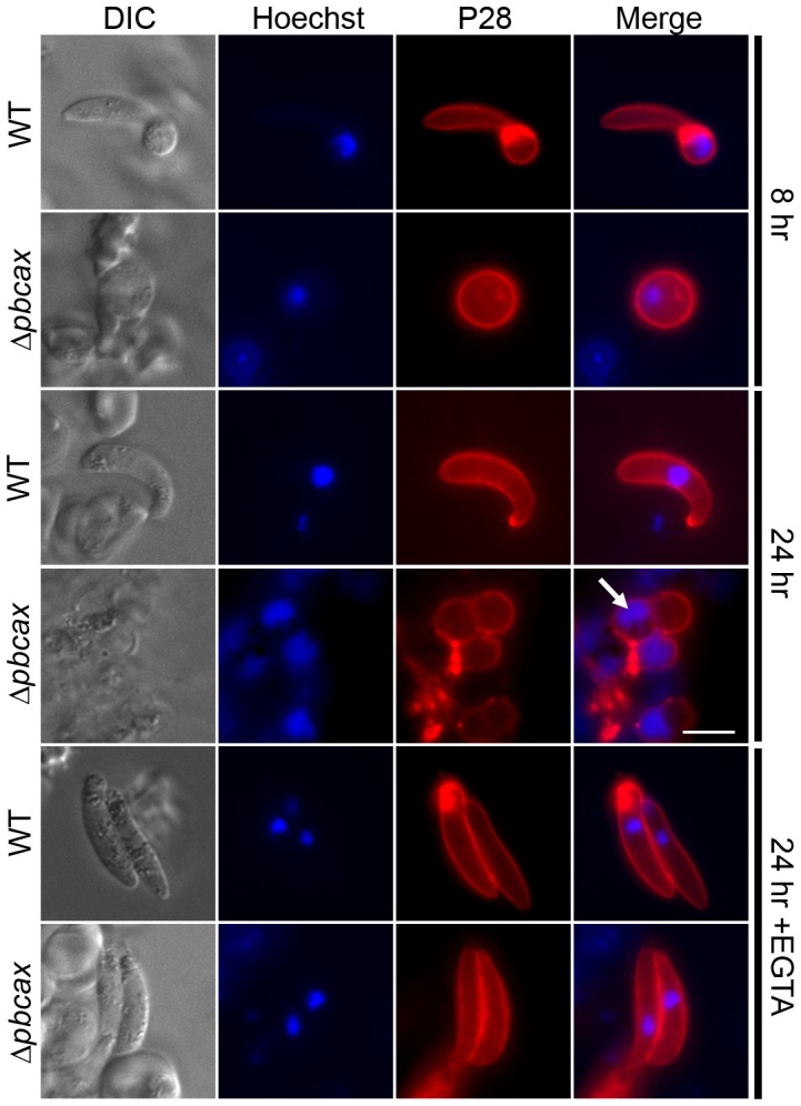
Immunofluorescence images of in vitro wild-type (WT) and Δpbcax cl9 P. berghei ookinete cultures, 8 and 24 h after gametocyte activation and in the presence of EGTA (10 mM; added directly prior to gametocyte activation). Parasites are immunostained for the female gamete/zygote/ookinete marker P28 (red) and co-stained with the nuclear marker Hoechst 33342 (blue). Development of elongated ookinetes was completely ablated in the Δpbcax cl9 line, a phenotype which could be reversed by the removal of extracellular Ca2+ using EGTA. The arrow indicates the diffuse DNA staining observed in Δpbcax cl9 parasites 24 h after gametocyte activation. Scale bar: 5 µm.