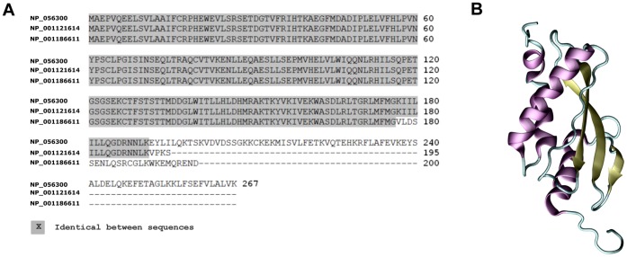
Figure 2. RSUME structural features.
A. BLAST multiple sequence alignment for RSUME267 (NP_056300), RSUME195 (NP_001121614) and RSUME200 (NP_001186611) isoforms. The identical aminoacids in the alignment are showed in gray. B. Ribbon Representation showing the secondary structure elements (alpha helixes are purple, beta-sheets are yellow and loops are cyan) of the RWD domain of RSUME obtained from the PDB (PDBid: 2EBK). The figure was made with the program VMD [29].

