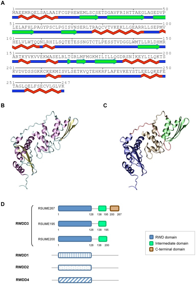
Figure 3. Domain characterization of RSUME.
A. Secondary structure prediction with PHYRE Server of the whole RSUME267 amino acid sequence. B. Ribbon representation of the RSUME267 structure as predicted by the PHYRE Server, showing the secondary structure elements: alpha helixes (purple), beta-sheets (yellow), loops (cyan). The Figure was made with the program VMD [29]. C. The same structure that in B but different colors encompassing the different regions of the RSUME267 protein. Blue residues 1 to 123, red residues 124 to 137, green residues 137 to 195, brown residues 196 to 267. D. Schematic comparison of the domains present in the three RSUME isoforms and in the RWD domain containing superfamily. Boxes, structural domains; lines, loops or unstructured regions; numbers, amino acid position.

