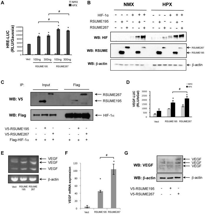
Figure 6. Effect of RSUME195 and RSUME267 over HIF-1 signaling pathway.
A. COS-7 cells were co-transfected with 500 ng of HRE-LUC report vector, 300 ng of Gaussia report vector and different concentrations of RSUME195 or 267 (100 or 300 ng) to evaluate their effect in HIF-1 transcriptional activity. Twenty-four hours after transfection cells were subjected to hypoxic conditions (1% O2, 5% CO2 and 94% N2) for 16 h. Then LUC activity was measured in the cell extracts. Each value was normalized to Gaussia value. Results are expressed as mean ± SEM from triplicates of one representative experiment of three experiments with similar results. *, p<0.05 compared with cells transfected with the empty vector (Vect) (ANOVA with Scheffè’s test). #, p<0.05 compared each concentration of cells, under hypoxia, transfected with RSUME195 vs. RSUME267 (ANOVA with Scheffè’s test). NMX, normoxia; HPX, hypoxia. B. COS-7 cells were co-transfected with 300 ng of Flag-HIF-1alpha and/or 500 ng of V5-RSUME195 or V5-RSUME267, or the corresponding empty vector (were both are absent). Twenty-four hours post-transfection, cells were subjected to hypoxic conditions (1% O2, 5% CO2 and 94% N2) for 16 h. Cell extracts were subjected to western blot. NMX, normoxia; HPX, hypoxia; Vect, cells transfected with the corresponding empty vectors. C. COS-7 cells were co-transfected with RSUME and HIF-1alpha. Twenty-four hours post-transfection, cells were subjected to hypoxic conditions (1% O2, 5% CO2 and 94% N2) for 16 h. RIPA cell extracts were inmunoprecipitated with anti-Flag antibody, and subjected to western blot with anti-V5 or anti-Flag antibodies. D. COS-7 cells were co-transfected with 500 ng of VEGF-LUC report vector, 300 ng of CMV-β Gal report vector and RSUME195 or 267. Twenty-four hours after transfection cells were subjected to hypoxic conditions (1% O2, 5% CO2 and 94% N2) for 16 h. Then LUC activity was measured in the cell extracts. Each value was normalized to β-galactosidase value. Results are expressed as mean ± SEM from triplicates of one representative experiment of three experiments with similar results. *, p<0.05 compared with cells transfected with the empty vector (Vect) under hypoxia (ANOVA with Scheffè’s test). #, p<0.05 compared the condition transfected with RSUME195 vs. RSUME267, under hypoxia (ANOVA with Scheffè’s test). NMX, normoxia; HPX, hypoxia. E. Semi-quantitative RT-PCR of endogenous VEGF and β-actin mRNA in HepG2 cells transfected with empty vector (Vect), RSUME195 or RSUME267, and subjected to hypoxia (1% O2, 5% CO2 and 94% N2) for 16 h, twenty-four hours after transfection. F. VEGF mRNA level was analyzed by quantitative real-time RT-PCR in triplicates in HeLa cell stimulated with hypoxia for 16 h, and the values are given as mean ± SEM after normalization to RPL19. *, p<0.05 compared with cells transfected with the empty vector (Vect) (ANOVA with Scheffè’s test). #, p<0.05 compared the condition transfected with RSUME195 vs. RSUME267 (ANOVA with Scheffè’s test). G. HepG2 cells were transfected with empty vector (Vect), RSUME195 or RSUME267, and subjected to hypoxia (1% O2, 5% CO2 and 94% N2) for 16 h, twenty-four hours after transfection. VEGF protein levels were analysed by western blot.

