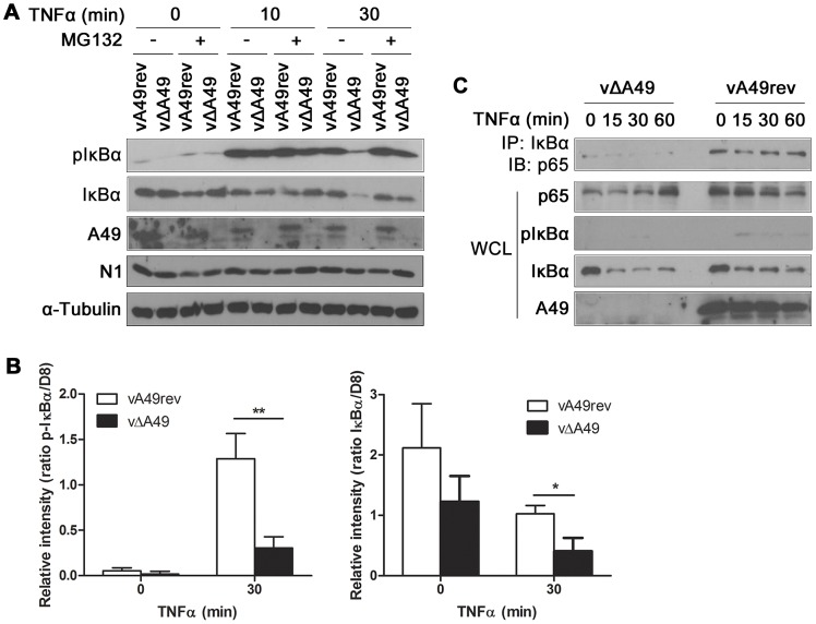
Figure 7. A49 interferes with IκBα degradation during viral infection.
(A) HeLa cells were infected with vA49rev or vΔA49 at 10 PFU/cell for 4 h, treated for 1 h with MG132 (20 µM) or vehicle only, and then stimulated with TNFα (200 ng/ml) as indicated. Cell extracts were separated by SDS-PAGE and analysed by immunoblotting with the antibodies indicated. N1 immunoblotting served as control for viral infection. (B) HeLa cells were infected and treated with TNFα in triplicate as in (A) and cell extracts were analysed by quantitative fluorescence immunoblotting. The amounts of p-IκBα and IκBα are shown as ratios compared with VACV protein D8. *p<0.05 or **p<0.01 comparing vA49rev with vΔA49. (C) HeLa cells were infected with vA49rev or vΔA49 at 10 PFU/cell for 6 h and then stimulated with TNFα (200 ng/ml) as indicated. Cells were then lysed in IP buffer and the lysates were immunoprecipitated with anti-IκBα antibody and immunoblotted for p65. In each assay, 2% of WCL of each sample was immunoblotted with the indicated antibodies.

