
Figure 2. Oxidized Residues Identified on the Stromally Exposed Regions of the D1 and D2 Proteins in the Vicinity of QA and PheoD1.
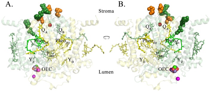
The T. vulcanus residues corresponding to the oxidatively modified spinach residues (Table 1) are highlighted. These oxidized residues are shown as spheres superimposed on monomer I of the T. vulcanus structure. For clarity, only the D1 and D2 proteins and their associated cofactors are shown. A. the view from outside Monomer I, looking towards the dimeric complex from within the plane of the membrane. B. the view from Monomer II looking towards its interface with Monomer I within the plane of the membrane. The D1 protein is shown in pale green and the D2 protein is shown in pale yellow. The oxidatively modified residues of D1 are shown in dark green while those of D2 are shown in orange. Various cofactors of both D1 and D2 are labeled and colored pale green or yellow, respectively. PheoD1 is shown in bright green. The non-heme iron is shown in bright red. The Mn4O5Ca cluster and its associated chloride ions are labeled as the OEC. Figs. 2–4 were produced using PYMOL [53].