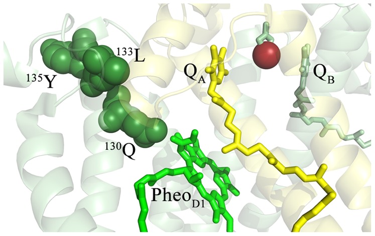
Figure 4. Detail of the Oxidized Residues in the Vicinity of PheoD1.

The T. vulcanus residues corresponding to the oxidatively modified spinach residues (Table 1) are highlighted and labeled. The D1 protein is shown in pale green and the D2 protein is shown in pale yellow. The oxidatively modified residues of D1 are shown in dark green, with the individual modified residues being labeled. PheoD1 is shown in bright green, QA is shown in yellow, QB in green and the non-heme iron is shown in bright red. For clarity, modified residues in the vicinity of QA (and detailed in Fig. 3) are not shown.
