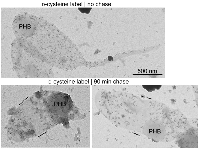
Figure 3. D-cysteine labeling of PG sacculi.
Transmission electron micrographs of d-Cys-labeled sacculi either before (top) or after (bottom) a 90-min chase period. These sacculi were obtained from cells grown in M2G medium. d-Cys residues were biotinylated and visualized by silver enhancement of nanogold-coupled anti-rabbit secondary antibody bound to anti-biotin primary antibody. Sacculi were counterstained with uranyl acetate. Dashed lines indicate areas of label clearing. PHB, polyhydroxybutyrate granules.

