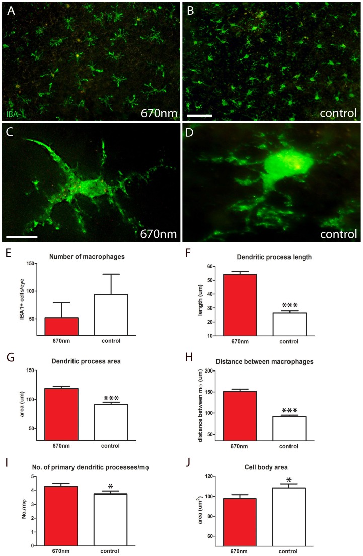
Figure 4. IBA-1 staining showed significantly different macrophage morphology between 670 nm and control groups.
A–D. RPE flat mounts labelled with IBA-1 (green) to identify macrophages. After 14 days of treatment with 670 nm light these cells had significantly altered morphology over a range of metrics. E. Number of IBA-1+ cells per eye was measured; there was a reduction in the number of macrophages following treatment, but this was not statistically significant. F,G. Dendritic process length and area were significantly increased by 670 nm light (27%, 28% respectively) following treatment (p = 0.0001 for each). H. The distance between macrophages was measured from nucleus of one cell to its nearest neighbour, which also showed a significant increase (p = 0.0001). I. Not only were the 670 nm treated cells larger they also had more primary processes (p = 0.05). J. Even though these cells had a greater dendritic field and territory they had smaller cell bodies in comparison to controls (p = 0.05). Abbreviations, retinal pigmented epithelial (RPE), ionized calcium-binding adaptor molecule 1 (IBA-1), macrophages (mφ). Scale bars A, B = 40 µm, C,D = 20 µm.

