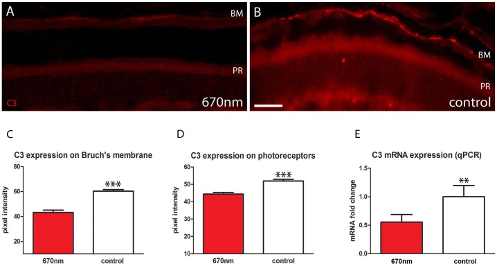
Figure 5. Outer retinal inflammation is significantly reduced following 670 nm treatment.
A.B. Retinal sections stained with C3 (red). This accumulates on Bruch’s membrane and outer segments. C.D Following 670 nm treatment C3 was significantly reduced on Bruch’s membrane and photoreceptor outer segments (p = 0.0001 for each). E. These data were confirmed with qPCR analysis, which showed again a statistically significant reduction in C3 expression following treatment (p = 0.0031). Abbreviations, Bruch’s membrane (BM), photoreceptor (PR), complement component (C3). Scale bars = 40 µm.

