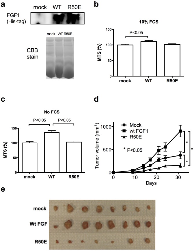
Figure 1. R50E suppresses tumorigenesis in vivo.
a. Transfected DLD-1 cells secrete WT FGF1 or R50E into culture medium. DLD-1 cells that stably express WT FGF1 or R50E were generated. The WT FGF1 and R50E have a 6His-tag at the N-terminus. To detect FGF1 secreted from the transfected cells, we analyzed the culture media by Western blotting with anti-6His antibodies. Mock-transfected cells were used as a control. As a loading control, we ran the same samples in gel in parallel and stained the gel with Coomassie Brilliant Blue (CBB). b. Proliferation of DLD-1 cells in the presence of 10% FCS. DLD-1 cells that secrete R50E grew in the medium that contains FCS in vitro at levels comparable to those of WT-FGF1 expressing cells or mock transfected cells. Statistical analysis was done by one-way ANOVA plus Tukey analysis. c. Proliferation of DLD-1 cells in the absence of FCS. DLD-1 cells that secrete R50E grew in vitro in the medium without FCS at levels comparable to that of mock-transfected cells. Cells that express WT FGF1 grew faster than mock-transfected and R50E expressing cells. Statistical analysis was done by one-way ANOVA plus Tukey analysis. d. The growth curve of DLD-1 cells in vivo. WT FGF1 enhanced tumor growth in vivo, while R50E suppressed it (as shown by the growth curve and the sizes of DLD-1 tumors removed at day 31). We injected the DLD-1 cells that secrete WT FGF1or R50E into nude mice (1 million cells/site) at right and left inguinal regions (4 mice per group, 2 tumors/mouse). Mock-transfected cells were used as a control. Statistical analysis of tumor sizes at Day 31 was done by t-test (n = 8 for mock and wt FGF, n = 7 for R50E). e. The sizes of tumors at Day 31. DLD-1 cells secreting wt FGF1 grew faster, and cells secreting R50E slower, than mock-transfected cells (n = 8 for mock and wt FGF, n = 7 for R50E).

