
Figure 2. R50E suppresses WT FGF1-induced endothelial cell migration.
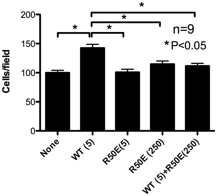
Lower side of the filter in the modified Boyden chamber was coated with fibronectin (10 µg/ml). The lower chamber was filled with serum-free medium with WT FGF1 (5 ng/ml) or the mixture of WT FGF1 and excess R50E (5 and 250 ng/ml, respectively). HUVECs were plated on the filter and incubated for 6 h. Chemotaxed cells were counted from the digital images of the stained cells. Data is shown as means +/− SE per field. Statistical analysis was done by one-way ANOVA plus Tukey analysis.