
Figure 4. Apoptosis is associated with induction of cleaved caspase-3 and cleaved caspase-7 in primary cortical neurons.
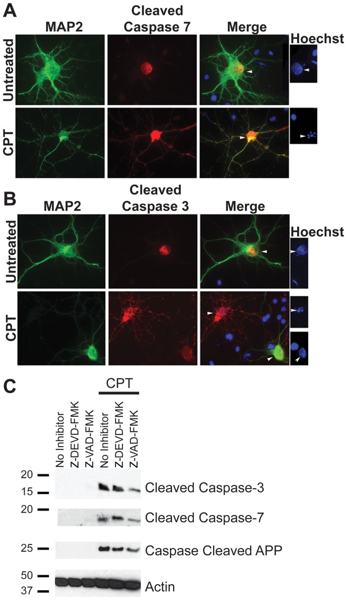
Primary neurons were cultured from E18 embryonic rat cortices for seven days, followed by treatment with 10 µM CPT to induce apoptosis. A and B show immunocytochemistry with MAP2 as a neuronal marker (green) and cleaved caspase-7 (A) or cleaved caspase-3 (B) antibodies (red) (Magnification: 63X). Hoechst staining, shown to the right of panels A and B, shows condensed or fragmented nuclei (indicative of apoptosis) in cells positive for cleaved caspases, whereas MAP2 positive neurons in the untreated samples show intact nuclei. (C) Western blot analysis of the lysates from neurons treated with CPT for 12 hours showed a significant induction in cleaved caspase-3 (top panel) and cleaved caspase-7 (second panel) levels. Activation of caspases was partially attenuated in the presence of 10 µM Z-DEVD-FMK and Z-VAD-FMK. Analysis of the lysates with an antibody generated against caspase-cleaved APP detected a single band of ∼25 kDa size. The level of this fragment was attenuated slightly in the presence of caspase inhibitors (Figure 4C, third panel). A reprobe of this blot using an antibody against β-Actin was performed as a control for protein loading (Figure 4C, bottom panel).