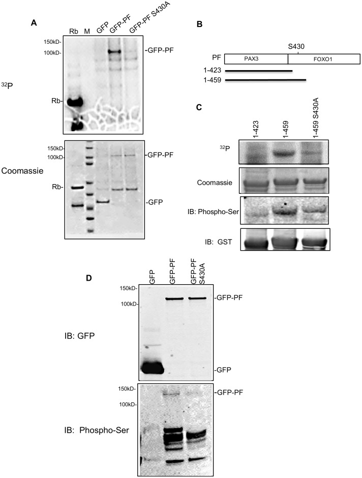
Figure 2. Cdk4 phosphorylates PAX3-FOXO1 at Ser430 in vitro and in vivo.
A) In vitro kinase assay using GFP or GFP-PAX3-FOXO1 (GFP-PF; wild-type or S430A mutant) pulled down from transfected Rh30 cells. Top panel: 32P Phospho-image. Bottom panel: protein substrates revealed by Coomassie blue staining. B) Schematic diagram of three GST-PF fusion proteins (showing only the PAX3-FOXO1 portion). PF, full length PAX3-FOXO1; 1–423, the N-terminal 423 residues of PAX3-FOXO1; 1–459, N-terminal 459 residues of PAX3-FOXO1; S430, Serine 430; PAX3, the PAX3 portion of PAX3-FOXO1; FOXO1, the FOXO1 portion of PAX3-FOXO1. C) In vitro kinase assay using bacterially expressed PAX3-FOXO1. From top to bottom panel: 32P Phospho-image (32P); protein substrates revealed by Coomassie blue staining (Coomassie); Western blot using phosphor-ser CDKs substrate antibody (IB: Phospho-Ser); and Western blot using anti-GST monoclonal antibody (IB: GST). D) Detection of phosphorylation using GFP or GFP-PF (wild-type or S430A mutant) proteins pulled down from transfected Rh30 cells. Top panel: Western blot using anti-GFP monoclonal antibody; Bottom panel: Western blot using phosphor-ser CDKs substrate antibody. Rb, positive control for Cdk4 activity; M, protein marker indicating molecular weight (kD).

