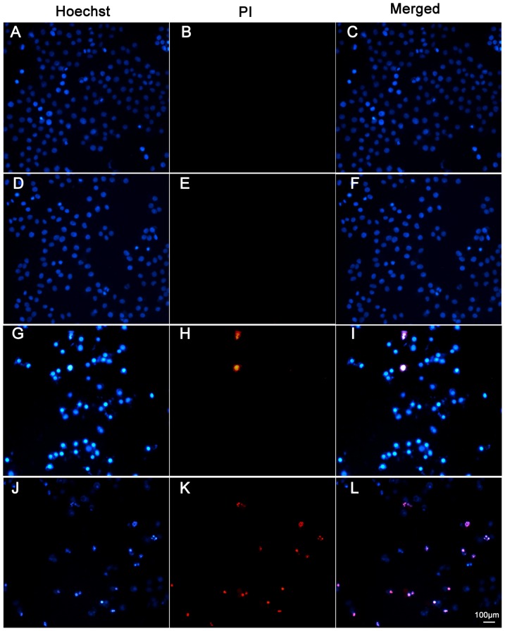
Figure 9. sch-fractions induce HepG2 apoptosis and necrosis.
HepG2 cells were treated in different sub-fractions and concentrations for 24 h, and then cell were stained with Hoechst/PI as mentioned in the ‘Materials and Methods’ section. (A–C) Cells were absence of any treatment; (D–F) Cells were exposure to 6.25 µg/ml sch of FRP; (G–I) Cells were exposure to 50 µg/ml sch of PSP; (J–L) Cells were exposure to 50 µg/ml sch of FRP; magnification:×200.

