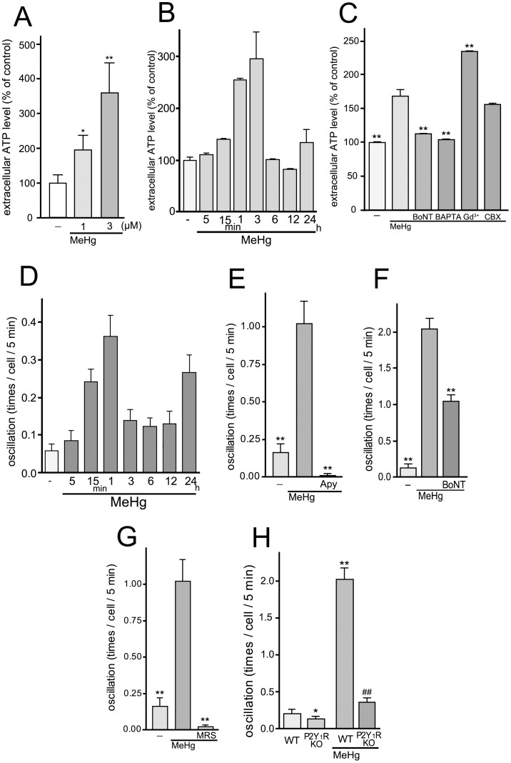
Figure 3. Exocytotic ATP release and Ca2+ oscillation in astrocytes evoked by MeHg.
(A) MeHg increased the extracellular ATP level of astrocytes. The effect of MeHg on the ATP release was concentration-dependent. MeHg was applied to the cell 15 min before the ATP measurement. *P<0.05 and **P<0.01 vs. control. (B) The time-course of extracellular ATP level of MeHg-treated astrocytes. MeHg (3 µM) increased extracellular ATP level of astrocytes, which was transient and peaked at 3 hr. (C) MeHg induces exocytotic ATP release from astrocytes. BoNT (5 units/ml, 24 hr pretreatment) and BAPTA-AM (10 µM) abolished MeHg (3 µM, 15 min)-induced ATP release from astrocytes. Gd3+(50 µM) rather enhanced ATP release, and CBX (100 µM) had no effect. **P<0.01 vs. MeHg. (D) The time-course of MeHg-induced changes in the frequency of Ca2+ oscillation. MeHg increased frequency of Ca2+ oscillation in astrocytes, which was transient and peaked at 1 hr. (E) MeHg increases the frequency of spontaneous calcium oscillations in astrocytes via enhancing ATP release. The number of Ca2+ oscillations was significantly increased by MeHg (3 µM, 30 min) and the increase was abolished by apyrase (Apy, 20 units/ml). **P<0.01 vs. MeHg. (F) Exocytotic pathway contributes to the MeHg-increased Ca2+ oscillation. BoNT (5 units/ml, 24 hr pretreatment) reduced the number of Ca2+ oscillations. **P<0.01 vs. MeHg. (G) Inhibitory effect of P2Y1 receptor antagonist on the Ca2+ oscillation frequency increased by MeHg. Ca2+ oscillation frequency was dramatically increased with MeHg (3 µM), and the increase was blocked by MRS2179 (MRS, 10 µM). **P<0.01 vs. MeHg. (H) P2Y1R is essential for the Ca2+ oscillation evoked by MeHg. WT astrocytes exhibited enhanced Ca2+ oscillation by MeHg (3 µM) but not in P2Y1R KO astrocytes. *P<0.05, **P<0.01 vs. control (WT), ##P<0.01 vs. MeHg (WT).

