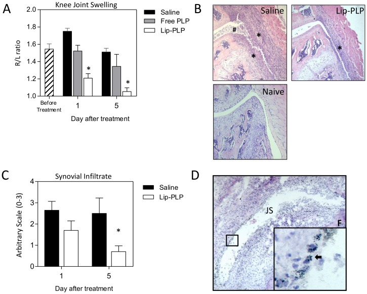
Figure 2. Liposomal targeting of PLP to the inflamed synovial lining strongly suppresses joint inflammation during AIA.
A: Knee joint swelling as measured by 99MTc-uptake is strongly suppressed after a single injection of Lip-PLP. B: Photomicrographs of frontal knee joint sections of mice with AIA at day 5 after treatment and naïve mice. Note that the inflammatory infiltrate is reduced in mice treated with Lip-PLP. Original magnification ×100, Asterisks points to synovial infiltrate, hash sign points to inflammatory exudates. C: Histological scoring of synovial infiltration at day 1 and day 5 after systemic treatment with Lip-PLP or saline. D: Silver staining of frontal knee joint sections of mice with AIA, treated by intravenous injection with gold-containing liposomes. Note that the silver staining of the gold particles is mostly observed within the synovial lining cells (arrows). Mice were treated at day 3 after induction of AIA. Values are the mean of 8 mice per group. Original magnification ×100; insert ×400. F = femur, JS = joint space. Statistical significance was determined by Student's t-test. * = P<0.05 compared to saline treatment.

