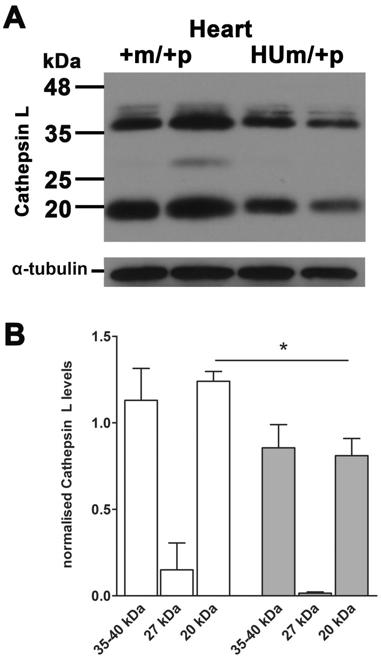
Figure 6. Embryonic heart Cathepsin L protein levels following maternal transmission of Igf2r+m/HUp .
(A) Western blots of Cathepsin L in mouse embryos at day E18.5. (B) Densitometry of protein levels normalised to α -tubulin loading control. Levels of cathepsin L were reduced in hearts of embryos with a humanised maternal allele (HUm) (P<0.05).

