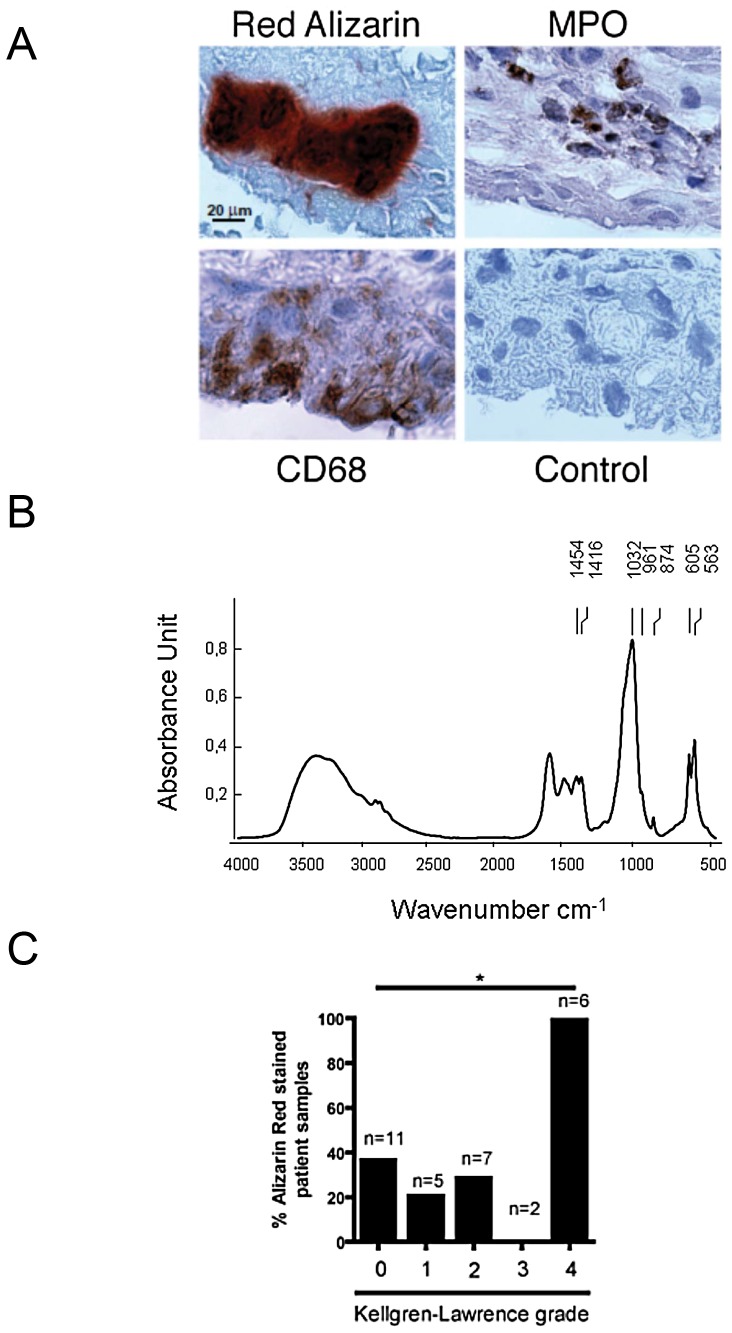
Figure 5. OA synovial membranes contained BCP crystals.
Consecutive paraffin sections of OA synoviums were analyzed with alizarin-red staining or by immunohistochemistry using anti-CD68 and anti-MPO primary antibodies (A). Biochemical composition of calcium-containing crystals in OA synovial membranes was assessed by FTIR spectroscopy, displaying a characteristic spectrum of Carbonated apatite (CA), with an absorption band peak at approximately 1035 cm-1 (B). Synovial membranes of different OA grades as assessed by Kellgren Lawrence score were harvested during arthroscopy. FTIR analysis showed the frequent presence of CA crystals in synovium membranes (C).

