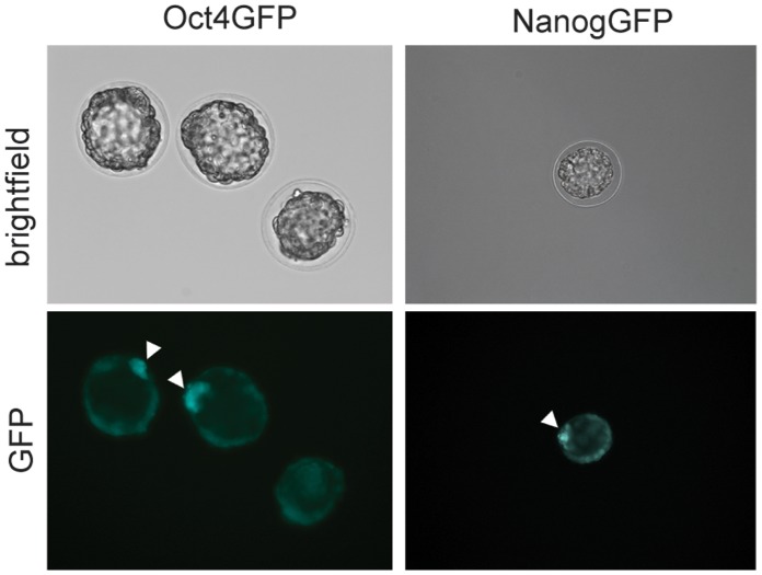
Figure 1. Oct4GFP and NanogGFP expression in inner cell mass of transgenic mouse embryos.
Blastocysts were isolated from pregnant Oct4GFP+ (left side) or NanogGFP+ (right side) mice. Bright field and fluorescence pictures were taken. Arrows indicate GFP+ cells in the inner cell mass (ICM) of the blastocysts.

