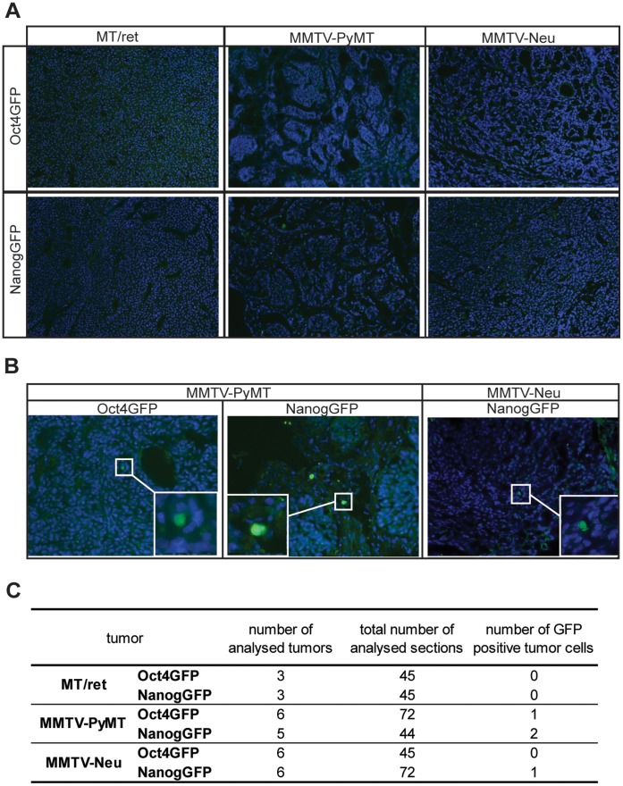
Figure 5. Only very rare GFP+ cells can be detected in compound MMTV-PyMT or MMTV-Neu tumors.
Sections of Oct4GFP+ or NanogGFP+ MT/ret, MMTV-PyMT and MMTV-Neu tumors were stained with an anti-GFP antibody and analysed for GFP positive cells. (A) Representative pictures of GFP immunofluorescence stainings showing the GFP negativity of virtually all sections from the compound tumors. (B) Pictures of the only GFP+ cells detected in single sections from Oct4GFP+ and NanogGFP+ MMTV-PyMT and MMTV-Neu tumors. The framed region is enlarged in the insert. (C) Table summarizing the immunofluorescence analysis, showing the number of analysed tumors per tumor model, the total number of analysed sections, and the total number of detected GFP+ cells per tumor model.

