
Figure 6. Spheroid cell culture does not induce Oct4GFP or NanogGFP expression in MMTV-PyMT or MMTV-Neu tumor cells.
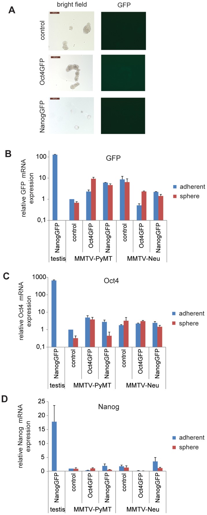
(A) No GFP+ cells can be detected in spheroids of Oct4GFP+ or NanogGFP+ MMTV-PyMT tumor cells. Oct4GFP+ or NanogGFP+ MMTV-PyMT tumor cells were freshly isolated from tumors, grown as spheres for one passage when pictures were taken. Representative bright field (left side) and fluorescent (right side) pictures are shown. Scale bar = 200 µm. (B-D) Freshly isolated Oct4GFP+ or NanogGFP+ MMTV-PyMT and MMTV-Neu tumor cells were either grown adherently (passage 1–4) or as spheres (passage 0) and analysed for relative expression of (B) GFP, (C) Oct4 and (D) Nanog mRNA by qPCR. mRNA expression of NanogGFP+ testis and compound MMTV-PyMT and MMTV-Neu tumor cells was compared to mRNA expression of control MMTV-PyMT tumor cells, which was set to 1. The analysis was performed in triplicate. The mean ± SEM is shown.