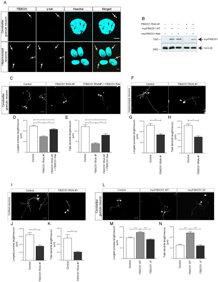
Figure 1. Centrosomal FBXO31 promotes axon and dendrite growth in neurons. A.
Cultured cerebellar granule neurons and hippocampal neurons were fixed using methanol followed by immunostaining with α-FBXO31 and α-γtubulin antibodies. The cells were counterstained with the DNA dye bisbenzimide Hoechst 33258. Arrows indicate centrosomes. Scale bar equals 5 µm. B. Cell lysates of HEK 293T cells transfected with indicated plasmids were probed with α-myc antibody. 14-3- ß served as a loading control. C. Representative images of cerebellar granule neurons transfected with empty control vectors, FXO31 RNAi #1 plasmid or FBXO31 RNAi #1 together with mycFBXO31-Res at DIV 0 and analyzed at DIV 4. Arrowheads indicate granule neuron cell bodies. Scale bar equals 50 µm. D. Quantification of longest process lengths of granule neurons shown in C (N = 3, n = 296, mean±SEM, one-way ANOVA *p<0.05, ***p<0.001). E. Quantification of total dendrite lengths of granule neurons shown in C (N = 3, n = 291, mean±SEM, one-way ANOVA, *p<0.05, ***p<0.001). F. Representative images of cultured hippocampal neurons transfected with control vector or FBXO31 RNAi #1 plasmids at DIV 1 and analyzed at DIV 5. Arrowheads indicate hippocampal neuron cell bodies. Scale bar equals 50 µm. G. Quantification of longest process lengths of hippocampal neurons shown in F (N = 3, n = 190, mean±SEM, unpaired t-test, ***p<0.001). H. Quantification of total dendrite lengths of hippocampal neurons shown in F (N = 3, n = 184, mean±SEM, unpaired t-test, **p<0.01). I. Representative images of cultured cortical neurons transfected with control vector or FBXO31 RNAi #1 plasmids at DIV 1 and analyzed at DIV 5. Arrowheads indicate cortical neuron cell bodies. Scale bar equals 50 µm. J. Quantification of longest process lengths of cortical neurons shown in I (N = 3, n = 164, mean±SEM, unpaired t-test, ***p<0.001). K. Quantification of total dendrite lengths of cortical neurons shown in I (N = 3, n = 147, mean±SEM, unpaired t-test, ***p<0.001). L. Representative images of cerebellar granule neurons transfected with empty control vector, mycFBXO31 wild type (WT) plasmid or mycFBXO31 ΔF mutant plasmid at DIV 0 and analyzed at DIV 3. Arrowheads indicate granule neuron cell bodies. Scale bar equals 50 µm. M. Quantification of longest process lengths of granule neurons shown in L (N = 3, n = 381, mean±SEM, one-way ANOVA ***p<0.001). N. Quantification of total dendrite lengths of granule neurons shown in L (N = 3, n = 341, mean±SEM, one-way ANOVA, ***p<0.001).

