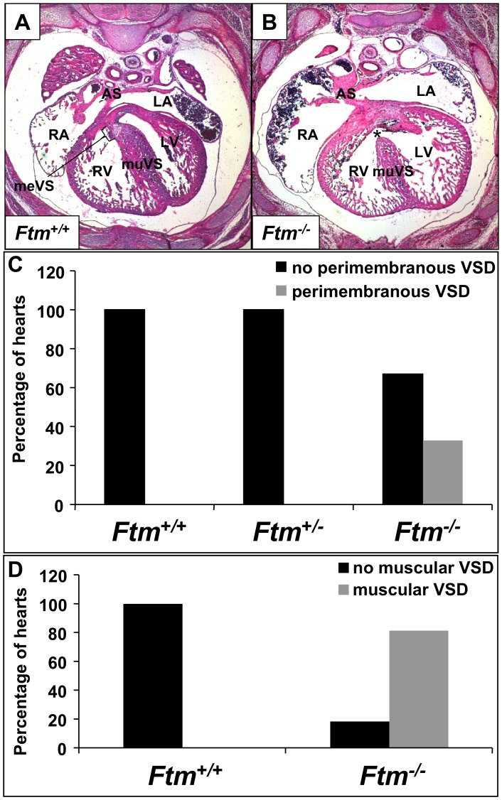
Figure 1. Ftm-deficient murine embryos show perimembranous and muscular ventricular septal defects.
(A, B) Hematoxylin and Eosin stainings at E14.5 on transverse heart sections. (A) In wild-type mouse embryos, the ventricular septum consists of a muscular part (muVS) and a membranous part (meVS). (B) In Ftm −/− embryos, the muscular VS displays a shorter and thinner shape and the membranous VS is missing (indicated by the asterisk) representing a perimembranous ventricular septal defect. (C) While in Ftm +/+ (n = 23) and Ftm +/− mice (n = 21) the heart develops normally, 33% of Ftm −/− mice (n = 27) show perimembranous ventricular septal defects. This statistics is based on investigations of mice at E13.5, E14.5, E15.5, E16.5 and E17.5. (D) Ftm +/+ mouse embryos (n = 23) do not suffer from muscular ventricular septal defects. 81.5% of all analyzed Ftm −/− embryos (n = 27) display muscular ventricular septal defects. Embryos at E13.5, E14.5, E15.5, E16.5 and E17.5 were examined in this context. LA, left atrium; RA, right atrium; LV, left ventricle; RV, right ventricle; meVS, membranous ventricular septum; muVS, muscular ventricular septum; AS, atrial septum.

