
Figure 3. Deformation of reconstructions and the effect of rim angle. A,
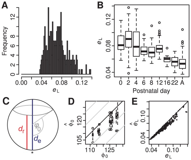
Histogram of the reconstruction error measure  obtained from 288 successfully reconstructed retinae. B, Relationship between deformation measure and age. “A” indicates adult animals. C, Schematic diagram of eye, indicating the measurements
  and
and  made on mouse eyes at different stages of development, and the rim angle
  derived from these measurements. Note that rim angle is measured from the optic pole (*). D, Rim colatitude
  that minimises reconstruction error versus the rim angle
  determined from eye measurements. Solid line shows equality and grey lines indicate ±10° and ±20° from equality. E, Minimum reconstruction error
  obtained by optimising rim angle versus reconstruction error
  obtained when using the rim angle from eye measurements. Solid line indicates equality.