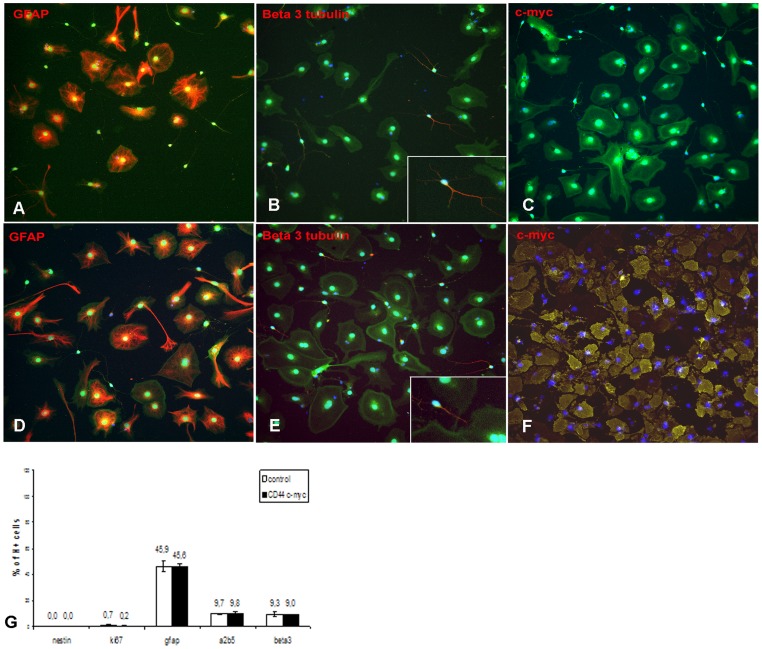
Figure 2. Transduction with lentiviral vector does not affect NPC differentiation in vitro.
NPCs were plated in the absence of EGF and FGF for 12 days. Immunocytochemistry for (A, D) GFAP, (B, E) ß3-tubulin, (C, F) c-myc of (A–C), control and (D–F), CD44-c-myc transduced actin eGFP cells (green). (G) Quantitative evaluation of the percentage of cells expressing the different markers over the total cell population identified by Hoechst staining (H+). Arrows in B and E point to cells enlarged in insets.

