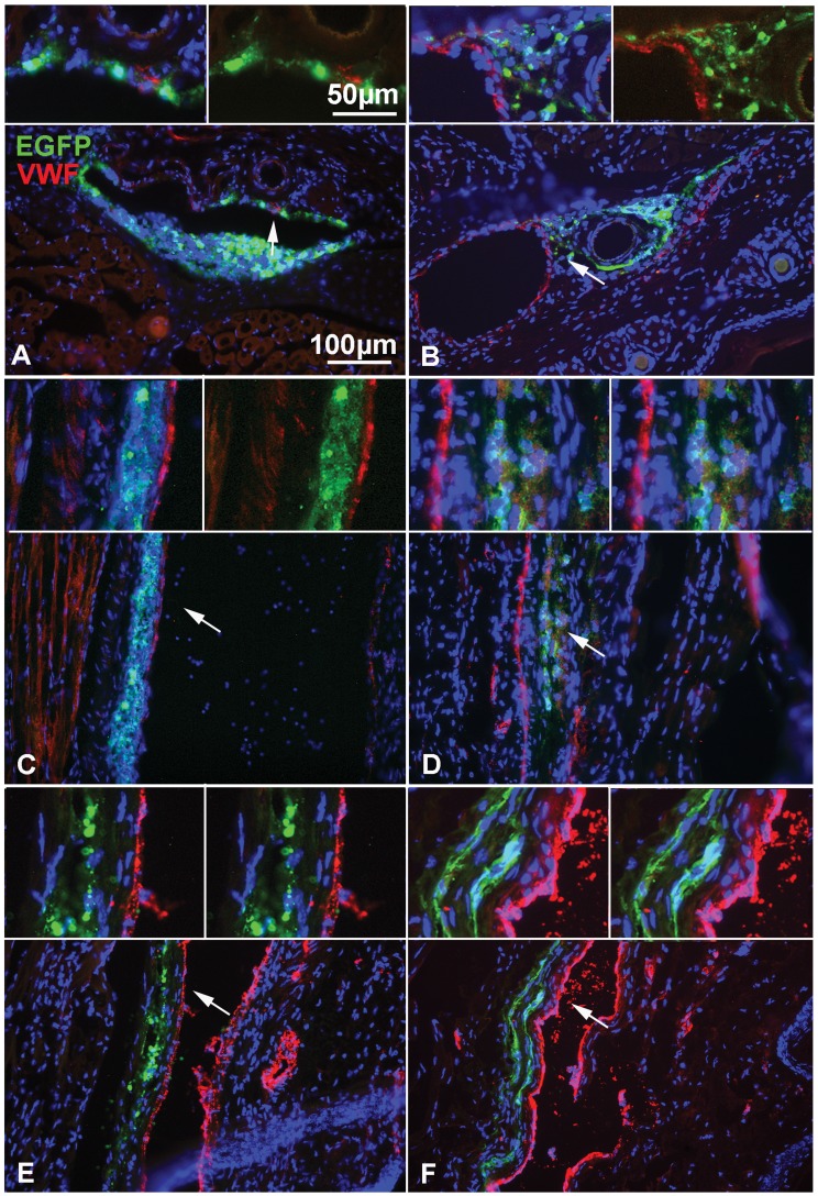
Figure 4. Time-course distribution of NPCs after i.v. delivery in the mouse tail.
(A–F) Detection of GFP(green) to localize grafted cells and of von Willebrand factor (VWF, red) to identify endothelial cells, reveals that NPCs are localized in or around the vascular wall at 6 h (A, B) and 12–24 h (C, D) post injection, but are more distant from the vascular wall at 21 days (E, F). (A, C, E) actin eGFP control-NPCs and (B, D, F) CD44-NPCs. CD44 NPCs become more rapidly elongated and distant from the vascular wall. Moreover, in D and F, GFP+ CD44-NPCs form a double wave, which is not observed in control NPCs. Arrows point to regions enlarged in insets on top of each panel.

