
FIG 4 .
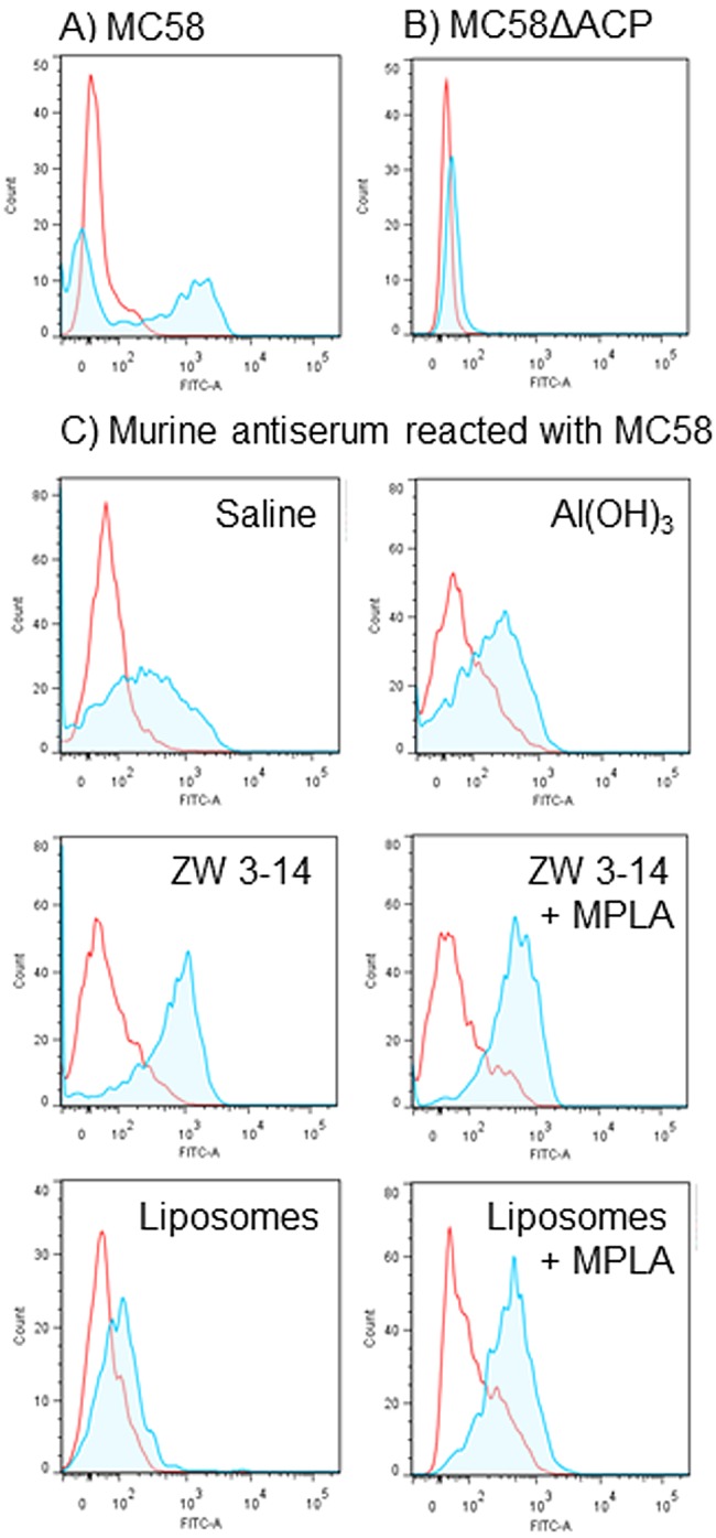
FACS analysis demonstrates expression of ACP on the surface of meningococci. (A and B) Reactivity of rabbit anti-rACP sera with MC58 and MC58 ∆ACP. The area within the red line shows the reactivity with preimmune rabbit sera (neat). The shaded area within the blue line shows the reactivity of rabbit anti-rACP sera (neat) with (A) MC58 (ACP+) and (B) MC58 ∆ACP (ACP−). (C) Reactivity of murine anti-rACP sera with MC58. Pooled murine antisera to rACP raised with the different adjuvants were reacted (1/10) with MC58. The areas within the red lines show the reactivity of sera from sham-immunized mice, and the shaded areas within the blue lines show the reactivity of sera from rACP-immunized mice.