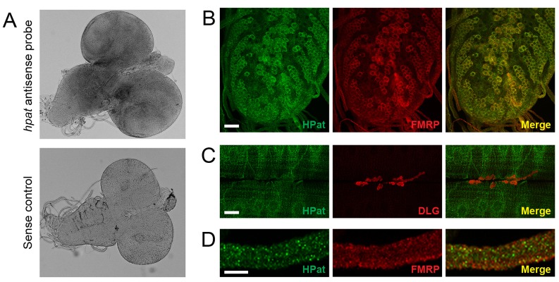
Fig. 2.

HPat is expressed in the larval nervous system. (A) Whole-mount RNA in situ hybridization of w1118 larval brains stained with either antisense or sense riboprobes directed against the hpat mRNA. (B) Larval ventral ganglion stained with rat antibodies against HPat (green) and/or FMRP (red) showing punctate cytoplasmic staining in the soma of most neurons. There is a significant amount of overlap between HPat and FMRP in the merged image. Scale bar: 20 µm. (C) NMJs double stained with antibodies against HPat (green) and postsynaptic Dlg (red). HPat exhibits punctate perinuclear localization in muscle but is not enriched at the NMJ. Scale bar: 20 µm. (D) Peripheral nerves exiting the ventral ganglion showing localization of both HPat (green) and FMRP (red). In the merged images in B and D, yellow spots indicate co-localization between HPat- and FMRP-containing punctae. Scale bar: 10 µm.
