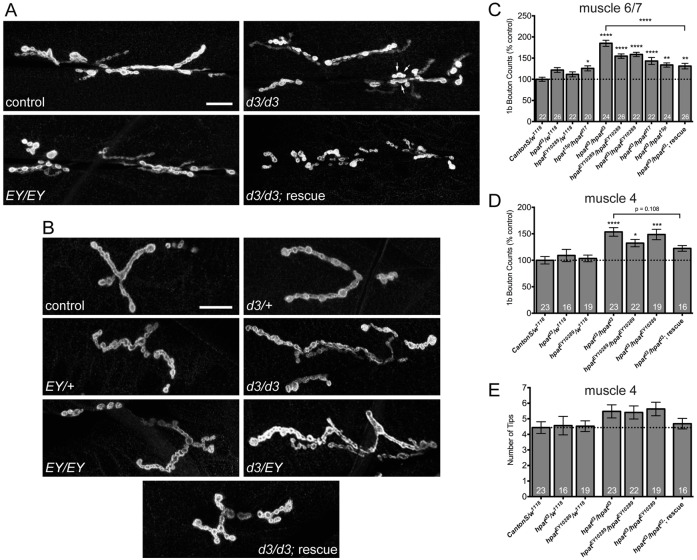
Fig. 3.
HPat negatively regulates synaptic terminal growth during larval development. (A) Representative images of muscle 6/7 and (B) muscle 4 NMJs from abdominal segment A3 labeled with an antibody against Dlg. Scale bar: 20 µm. Arrows in the hpatd3/hpatd3 panel in A indicate a cluster of terminal synaptic boutons. (C) Quantification of the number of type 1b synaptic boutons at muscle 6/7 and (D) muscle 4 synaptic clefts. (E) Quantification of the number of total bouton branch tips at muscle 4 for each indicated genotype. Unless otherwise indicated, stars denote statistical significance compared to Canton-S/w1118 controls (*P<0.05; **P<0.01; ***P<0.001; ****P<0.0001; one-way ANOVA; Tukey’s post-hoc test).

