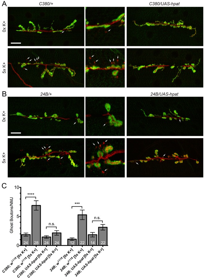
Fig. 6.
Neuronal HPat negatively regulates activity-dependent synaptic terminal growth. (A,B) Representative images of muscle 6/7 NMJs from abdominal segment A3 labeled with antibodies against presynaptic HRP (red) and postsynaptic Dlg (green). HPat expression was driven pre- or postsynaptically using the C380-Gal4 and 24B-Gal4 drivers. Arrows indicate the presence of immature ghost boutons (HRP+ Dlg−). Middle panels are magnified images of regions indicated by arrows in 0×K+ and 5×K+ treatment groups. Stimulated (5×K+) larvae of each genotype were taken through a spaced high-K+ paradigm. Unstimulated (0×K+) larvae were taken through a low K+ pseudo-stimulation paradigm. Scale bars: 20 µm. (C) Quantification of the number of ghost boutons per NMJ at muscle 6/7 clefts. Asterisks indicate statistical significance between the indicated groups (***P<0.001; ****P<0.0001; Kruskal–Wallis; Dunn’s post-hoc test). Note the a 3- to 4-fold increase in the number of ghost boutons is observed at control NMJs following high-K+ depolarization. This increase is almost completely suppressed by both pre- and postsynaptic overexpression of HPat (down to a 1.4- and 1.5-fold increase, respectively). n.s., not significant.

