Figure 6.
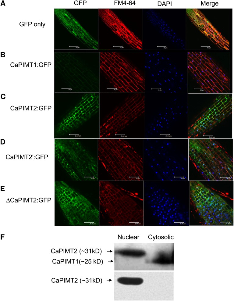
Subcellular localization of CaPIMT1, CaPIMT2, CaPIMT2′, and ΔCaPIMT2 (CaPIMT2 lacking the 56-amino acid N terminus). A to E, Localization of GFP, CaPIMT1:GFP, CaPIMT2:GFP, CaPIMT2′:GFP, and ΔCaPIMT2:GFP in roots of Arabidopsis transgenic plants. Fluorescence images were taken using a confocal-laser scanning microscope. The plasma membrane and nuclei were stained by FM4-64 and 4′,6-diamino-phenylindole (DAPI), respectively. F, Western blot of nuclear and cytosolic proteins of 24-h-imbibed chickpea seed. Approximately 50 μg of nuclear or cytosolic protein was separated by 12% SDS-PAGE and probed with anti-PIMT (top panel) or anti-CaPIMT2 (bottom panel) antibody.

