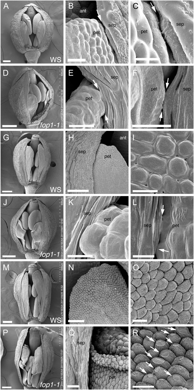
Figure 2.
Scanning electron microscopy images of floral organ surface. A to C, G to I, and M to O, Wild type. D to F, J to L, and P to R, fop1-1. A to F, Stage 10. G to L, Stage 11. M to R, Stage 12. B and C, Higher magnification of A, showing a space between petal and sepal (arrows). E and F, Higher magnification of D, showing direct contact between petal and sepal (arrows). H and I, Higher magnification of G, showing straight petal elongation (G) and abaxial epidermis of petals (I) starting the deposition of the epicuticular nanoridges on their surface. K and L, Higher magnification of J, showing direct contact of petal and sepal (arrows). N and O, Petal surface of the same flower as M. Abaxial epidermal cells of petals deposit nanoridges on cell surface. Q, Higher magnification of P, showing the petal edge turning outward. R, Higher magnification of petals in Q, showing the trace of friction (arrows). The front side sepal is removed in A, D, G, J, M, and P to show the inside of flowers. sep, Sepal; pet, petal; ant, anther. Bars = 200 μm (D, G, J, M, and P), 100 μm (A and H), 50 μm (N), 20 μm (Q), 10 μm (B, E, I, O, and R), 5 μm (C, K, and L), and 3 μm (F).

