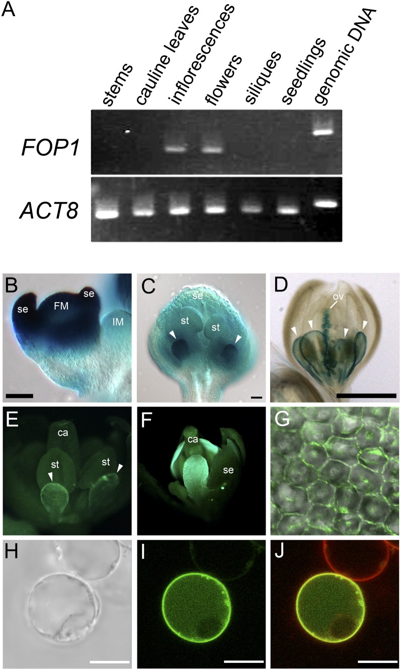
Figure 4.
FOP1 is expressed in elongating petals. A, RT-PCR analysis of FOP1 transcripts. FOP1 is expressed in inflorescences, including young floral buds, and in open flowers. ACT8 was used as a control. B to D, GUS expression in inflorescences and flowers carrying the FOP1p:GUS transgene. B, GUS is expressed strongly in floral meristem and sepal primordia. C, Strong GUS expression in petal primordia of a stage 9 flower (arrowheads). D, GUS expression is observed at petal margin (arrowheads) and ovules in a stage 11 flower. E to G, GFP fluorescence in fop1-1 flowers of plants carrying the FOP1p:FOP1-GFP transgene. The fusion protein is expressed preferentially at the margin of petals of a stage 10 flower (E) and in the apical part of petals of a stage 12 flower (F). G, A merged image of bright-field and GFP images of petal epidermal cells. The FOP1-GFP fusion protein is localized at the cell periphery. H to J, Suspension cell expressing GFP-FOP1 transiently. H, Bright-field image. I, GFP image. J, Merged image of the GFP and FM4-64 images, showing that GFP-FOP1 is localized to plasma membrane. Note that the upper cell, which is supposed not to carry the construct, shows only FM4-64 signal. IM, Inflorescence meristem; FM, floral meristem; se, sepal; st, stamen; ov, ovule; ca, carpel. Bars = 50 μm (B and C), 500 μm (D), and 10 μm (H–J).

