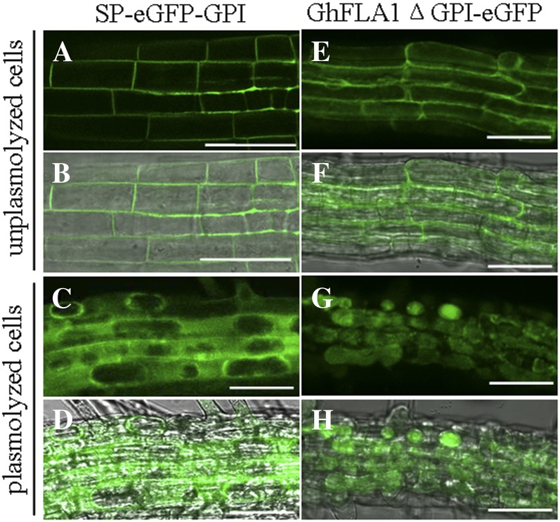
Figure 5.
Subcellular localization of GhFLA1 protein in root cells of 7-d-old Arabidopsis transgenic seedlings. A to D, Transgenic root cells expressing GhFLA1(SP)-eGFP-GhFLA1(GPI). E to H, Transgenic root cells expressing GhFLA1(noGPI)-eGFP. A, C, E, and G are GFP fluorescence images, and B, D, F, and H are the fluorescence images merged with their respective bright-field images. A, B, E, and F, Unplasmolyzed root cells. C, D, G, and H, Root cells plasmolyzed with 4% NaCl. Bars = 50 µm. [See online article for color version of this figure.]

