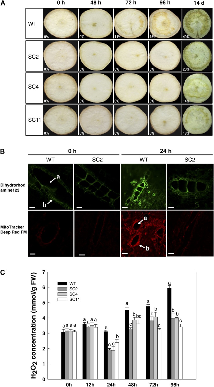
Figure 5.
Delayed PPD occurrence of storage roots from transgenic cassava by visual and fluorescence determination. A, Visual detection of PPD occurrence using the International Center for Tropical Agriculture method (Wheatley et al., 1985; Morante et al., 2010). The levels of vascular discoloration are indicated by percentages using ImageJ processing software. B, Inhibition of mitochondrial ROS generation during the PPD process in transgenic storage roots detected by fluorescent probes. The fluorescence intensity of oxidized rhodamine was observed with a fluorescence microscope (Zeiss LSM 510 META) with excitation/emission of 488/515 nm and 635/680 nm for DHR and MitoTracker-Deep Red FM, respectively. a, Xylem vessel; b, bundle sheath. Bars = 20 μm. C, Changes of H2O2 concentrations during PPD between wild-type (WT) and transgenic cassava. Values labeled with different letters (a, b, and c) at the same time point are significantly different by Duncan’s multiple comparison tests at P < 0.05. FW, Fresh weight. [See online article for color version of this figure.]

