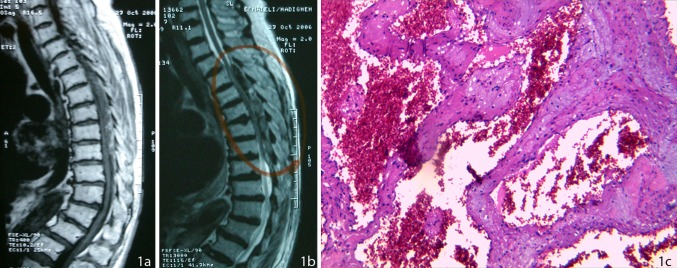
Fig. 1.
a Sagittal T1W MRI of the thoracic spine showing a heterogeneous epidural mass compressing the spinal cord at T5–T9 level, b sagittal T2W MRI showing the heterogeneous epidural lesion compressing the spinal cord at the same level, and c dilated capillaries with a thin wall and a simple endothelial lamina with thin adventitia. Fibrous scar tissue with scattered hemosiderin calcification located in between the vascular spaces

