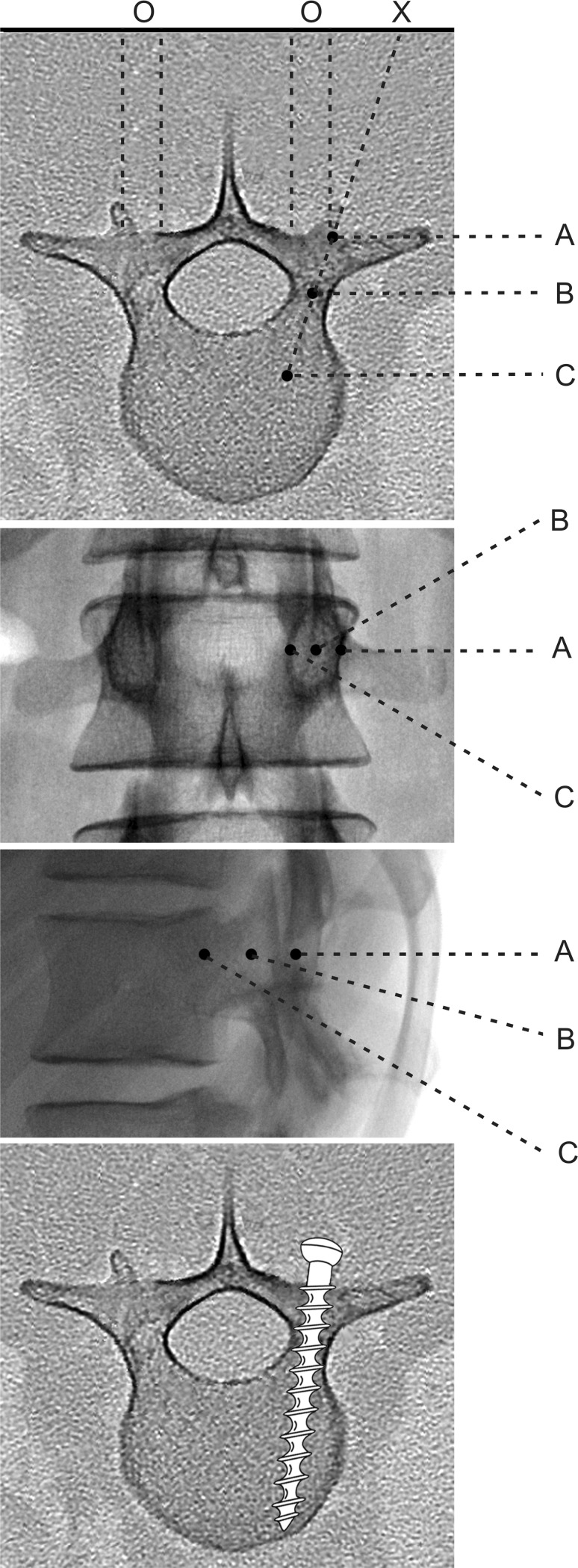
Fig. 4.
Diagrams illustrating the anatomical principles of percutaneous pedicle screw insertion: axial view, a.p. view, lateral view, and axial view (top to bottom). 0: Projection point of the pedicle onto the backskin. X: Incicion point. First, the needle tip is docked onto the lateral margin of the pedicle in three (right pedicle) and nine (left pedicle) o’clock position (position A). As the needle advances toward the base of the pedicle on the lateral image, it should approach the pedicle center on the a.p. image (position B). When the Jamshidi needle exceeds the posterior wall of the vertebra on the lateral image, the tip of the needle has to remain lateral to the medial pedicle wall on the a.p. image (position C)

