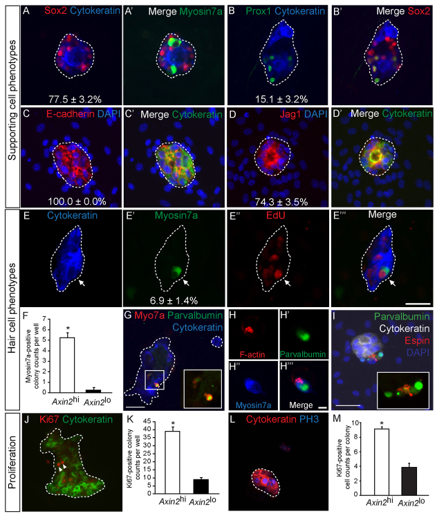
Fig. 5.
Purified Axin2hi cells acquire hair cell and supporting cell phenotypes. (A-D′) Axin2hi cells formed colonies uniformly expressed cytokeratin and E-cadherin, whereas 77.5±3.2%, 74.3±3.5% and 15.1±3.2% of all cytokeratin-positive colonies expressed Sox2, jagged 1 and Prox1, respectively (n=3 for each, median=22 Sox2-positive cells, 22 jagged 1-positive cells and 4 Prox1-positive cells per colony). (E-E″′) In the presence of EdU, myosin 7a-positive, EdU-positive hair cell-like cells were observed on cytokeratin-positive colonies (arrow) (n=4). (F) Myosin 7a-positive hair cells were observed more frequently among Axin2hi (median indicates one myosin 7a-positive cell per colony) than Axin2lo colonies (P<0.005, n=4). (G) All myosin 7a-positive hair cell-like cells also expressed parvalbumin. (H-H″′) A polarized pattern of filamentous actin on the hair cell-like cells was observed. (I) Two parvalbumin-positive hair cell-like cells with espin expression localized to one pole of the cell. (J,K) Cytokeratin-positive colonies containing actively proliferating Ki67-labeled cells (arrowheads) were noted more commonly in Axin2hi than in the Axin2lo colonies (P<0.001, n=3). (L) Within each Axin2hi colony, a subset of cells expressed the M-phase marker phosphohistone 3 (PH3). (M) There were more Ki67-positive cells in Axin2hi than Axin2lo colonies (P<0.0001, n=3). Scale bars: 25 μm in A-E″′,I,L; 50 μm in G,J; 5 μm in H-H″′. Data are mean±s.d. Asterisks indicate statistical significance.

